Category Books
- Fiction Books & Literature
- Graphic Novels
- Horror
- Mystery & Crime
- Poetry
- Romance Books
- Science Fiction & Fantasy
- Thrillers
- Westerns
- Ages 0-2
- Ages 3-5
- Ages 6-8
- Ages 9-12
- Teens
- Children's Books
- African Americans
- Antiques & Collectibles
- Art, Architecture & Photography
- Bibles & Bible Studies
- Biography
- Business Books
- Christianity
- Computer Books & Technology Books
- Cookbooks, Food & Wine
- Crafts & Hobbies Books
- Education & Teaching
- Engineering
- Entertainment
- Foreign Languages
- Game Books
- Gay & Lesbian
- Health Books, Diet & Fitness Books
- History
- Home & Garden
- Humor Books
- Judaism & Judaica
- Law
- Medical Books
- New Age & Spirituality
- Nonfiction
- Parenting & Family
- Pets
- Philosophy
- Political Books & Current Events Books
- Psychology & Psychotherapy
- Reference
- Religion Books
- Science & Nature
- Self Improvement
- Sex & Relationships
- Social Sciences
- Sports & Adventure
- Study Guides & Test Prep
- Travel
- True Crime
- Weddings
- Women's Studies
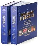
Diagnostic Ultrasound: 2-Volume Set » (3rd Edition)

Authors: Carol M. Rumack, Stephanie R. Wilson, J. William Charboneau, Jo-Ann Johnson
ISBN-13: 9780323020237, ISBN-10: 0323020232
Format: Hardcover
Publisher: Elsevier Health Sciences
Date Published: November 2004
Edition: 3rd Edition
Author Biography: Carol M. Rumack
Book Synopsis
The third edition has been substantially revised, with many new images (there are now more than 5000). New sections have been added on new imaging techniques, a new chapter has been added on organ transplantation, and obstetrics and gynecology receives greatly expanded treatment. The two volumes contain 62 chapters, divided into the parts of the body. The second volume is devoted entirely to obstetric, fetal, and pediatric sonography, with chapters on the safe use of ultrasound in obstetrics, structural anomalies in the first trimester, sonographic markers of fetal chromosomal defects, sonographic evaluation of the placenta, invasive fetal procedures, neonatal and infant brain imaging, and the pediatric spinal canal. Among the chapter title s in volume one are intraoperative and laparoscopic sonography of the abdomen, and sonography of the thyroid gland, the peripheral veins, the rotator cuff, and the tendons. Initial chapters are devoted to the physics of ultrasound. The contributors an d editors are specialists at hospitals and medical schools mainly in the US and Canada; some are in Europe. Annotation ©2004 Book News, Inc., Portland, OR
Paula J. Woodward
This is the second edition of this two-volume comprehensive textbook on principles, imaging, and diagnosis by sonography. The first edition was released at the RSNA in 1991. The technology, imaging accuracy, and potential applications of ultrasound have changed dramatically in the last six years. The purpose of this book is to build on the framework of the first edition and include the latest state-of-the-art concepts including color and power Doppler, hysterosonography, laparoscopic sonography, and ultrasound guided interventional procedures. This book can serve as a reference for practicing radiologists. In addition its organization and excellent illustrations make it particularly good for sonographers and residents just beginning to learn ultrasound. There are many excellent line drawings included within each chapter which complement the text. The ultrasounds are all of the highest quality and present the pathology well. New in this edition are colored enhanced boxes which highlight important monographic findings or differential diagnoses. Key terms and concepts are in bold face facilitating rapid referencing. The first edition of this book is one of the most commonly used textbooks in the field of ultrasound. This new edition is approximately 25 percent larger with marked expansion in obstetrics and gynecology. The majority of the images have been updated from the prior edition to reflect state-of-the-art techniques. The organizational changes have made it even easier to use as a reference text. This represents a significant improvement in what was already one of the most comprehensive reference books available on diagnostic ultrasound.
Table of Contents
VOLUME ONE
I. PHYSICS
1. Physics of Ultrasound
2. Biologic Effects and Safety
3. Microbubble Contrast Agents for Ultrasound Imaging: Where, How and Why?
II. ABDOMINAL, PELVIC AND THORACIC SONOGRAPHY
4. The Liver
5. The Spleen
6. The Biliary Tract and Gallbladder
7. The Pancreas
8. The Gastrointestinal Tract
9. The Urinary Tract
10. The Prostrate
11. The Adrenal Glands
12. The Retroperitoneum and Great Vessels
13. The Abdominal Wall
14. The Peritoneum
15. Gynecologic Ultrasound
16. Gestational Trophoblastic Disease
17. The Thorax
18. Ultrasound-Guided Biopsy and Drainage of the Abdomen and Pelvis
19. Organ Transplantation
III. INTRAOPERATIVE SONOGRAPHY
20. Intraoperative and Laparoscopic Sonography of the Abdomen
IV. SMALL PARTS, CAROTID ARTERY, AND PERIPHERAL VESSEL SONOGRAPHY
21. The Thyroid Gland
22. The Parathyroid Glands
23. The Breast
24. The Scrotum
25. The Rotator Cuff
26. The Tendons
27. The Extracranial Cerebral Vessels
28. The Peripheral Arteries
29. The Peripheral Veins
VOLUME TWO
V. OBSTETRIC AND FETAL SONOGRAPHY
30. Overview of Obstetric Sonography
31. The Prudent and Safe Use of Ultrasound in Obstetrics
32. The First Trimester
33. Structural Anomalies in the First Trimester
34. Sonographic Markers of Fetal Chromosomal Defects
35. Sonography of Multifetal Pregnancy
36. The Fetal Face and Neck
37. The Fetal Head and Brain
38. The Fetal Spine
39. The Fetal Chest
40. The Fetal Heart
41. The Fetal Abdominal
42. The Fetal Urogenital Tract
43. The Fetal Musculoskeletal System
44. Fetal Hydrops
45. Fetal Measurements—Normal and Abnormal Fetal Growth
46. Biophysical Profile Scoring
47. Doppler Assessment of Pregnancy
48. Sonographic Evaluation of the Placenta
49. Cervical Ultrasound and Preterm Labor
50. Invasive Fetal Procedures
VI. PEDIATRIC SONOGRAPHY
51. Neonatal and Infant Brain Imaging
52. Doppler of the Neonatal and Infant Brain
53. Doppler of the Brain in Children
54. Pediatric Head and Neck Masses
55. The Pediatric Spinal Canal
56. The Pediatric Chest
57. The Pediatric Liver and Spleen
58. The Pediatric Kidney and Adrenal Glands
59. The Pediatric Gastrointestinal Tract
60. The Pediatric Pelvic Sonography
61. Pediatric Musculoskeletal Ultrasound
62. Pediatric Interventional Sonography
Subjects

