Category Books
- Fiction Books & Literature
- Graphic Novels
- Horror
- Mystery & Crime
- Poetry
- Romance Books
- Science Fiction & Fantasy
- Thrillers
- Westerns
- Ages 0-2
- Ages 3-5
- Ages 6-8
- Ages 9-12
- Teens
- Children's Books
- African Americans
- Antiques & Collectibles
- Art, Architecture & Photography
- Bibles & Bible Studies
- Biography
- Business Books
- Christianity
- Computer Books & Technology Books
- Cookbooks, Food & Wine
- Crafts & Hobbies Books
- Education & Teaching
- Engineering
- Entertainment
- Foreign Languages
- Game Books
- Gay & Lesbian
- Health Books, Diet & Fitness Books
- History
- Home & Garden
- Humor Books
- Judaism & Judaica
- Law
- Medical Books
- New Age & Spirituality
- Nonfiction
- Parenting & Family
- Pets
- Philosophy
- Political Books & Current Events Books
- Psychology & Psychotherapy
- Reference
- Religion Books
- Science & Nature
- Self Improvement
- Sex & Relationships
- Social Sciences
- Sports & Adventure
- Study Guides & Test Prep
- Travel
- True Crime
- Weddings
- Women's Studies
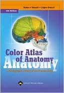
Color Atlas of Anatomy: A Photographic Study of the Human Body » (6th Edition)

Authors: Johannes W. Rohen, Chihiro Yokochi, Elke Lutjen-Drecoll
ISBN-13: 9780781790130, ISBN-10: 0781790131
Format: Hardcover
Publisher: Lippincott Williams & Wilkins
Date Published: May 2006
Edition: 6th Edition
Author Biography: Johannes W. Rohen
Rohen, Johannes W., Drmed, Drmedhc (Univ of Erlangen-Nurnberg);
Yokochi, Chihiro, MD (Kanagawa Dental Coll, Yokosuka); and: Elke Lütjen-Drecoll, MD; Lynn J. Romrell, PhD.
Book Synopsis
This atlas features outstanding full-color photographs of actual cadaver dissections, with accompanying schematic drawings and diagnostic images. The photographs depict anatomic structures more realistically than illustrations in traditional atlases and show students exactly what they will see in the dissection lab.
Chapters are organized by region in order of a typical dissection. Each chapter presents structures both in a systemic manner from deep to surface, and in a regional manner.
This edition has sixteen additional pages of clinical images—including CT and MRI—that students can compare with cross-sectional anatomic photographs. Many pictures have been electronically enhanced or rescanned for better contrasts.
Larry R. Cochard
This is the third edition of an anatomic atlas combining photographs of human cadaver dissections with didactic line drawings, CT scans, and MR images. Like most photographic atlases, it is intended to depict human anatomy in the most realistic manner possible for use in dissection labs as an aid to identification, for lab review, or in anatomy classes where cadavers are in short supply. Although the book is primarily intended for first-year medical students, it will be a good reference for senior students, residents, or anyone else learning or reviewing anatomy. Most of the atlas consists of high quality color photos of sequential dissections organized by body regions. The pictures have unobtrusive, numbered leader lines with with keyed structures listed on each page. Unique features that make this atlas more than just an aid to cadaver identification are imaging sections and colored line drawings. The latter include nerve and vessel schemes, topography, systemic overviews, muscle functions, and other anatomic points that are helpful in learning anatomy. The book is attractive, comprehensive, and has an extensive index. Three editions have resulted in a good product, but the process has been mostly fine-tuning instead of a significant change of content. It takes a little while to find changes from the second edition. Some photos have been replaced, a few line drawings have been added, and most new MR images are of the extremities. As a learning tool, this atlas has the best of the old and the new. The didactic figures found in traditional atlases, the imaging modalities, and the excellent pictures combine to make this the most versatile of the photographic atlases. If you have a copy ofthe second edition, though, hang on to it.
Table of Contents
| 1 | General Anatomy | 1 |
| Organization of the Human Body | 2 | |
| Skeleton of the Human Body | 4 | |
| Ossification of the Bones | 6 | |
| Bone Structure | 8 | |
| Joints | 10 | |
| Muscles and Tendon Attachments | 13 | |
| Organization of the Nervous System | 14 | |
| Organization of the Circulatory System | 16 | |
| Organization of the Lymphatic System | 18 | |
| 2 | Head and Neck | 19 |
| Bones of the Skull | 20 | |
| Disarticulated Skull | 24 | |
| Sphenoidal and Occipital Bones | 24 | |
| Temporal Bone | 26 | |
| Frontal Bone | 28 | |
| Calvaria | 29 | |
| Base of the Skull | 30 | |
| Skull of the Newborn | 35 | |
| Median Section through the Skull | 36 | |
| Disarticulated Skull | 38 | |
| Ethmoidal Bone | 38 | |
| Ethmoidal and Palatine Bones | 39 | |
| Palatine Bone and Maxilla | 40 | |
| Maxilla, Zygomatic Bone, and Bony Palate | 45 | |
| Orbit, and Nasal and Lacrimal Bones | 47 | |
| Bones of the Nasal Cavity | 48 | |
| Septum and Cartilages of the Nose | 49 | |
| Maxilla and Mandible with Teeth | 50 | |
| Deciduous and Permanent Teeth | 51 | |
| Mandible and Dental Arch | 52 | |
| Ligaments of the Temporomandibular Joint | 53 | |
| Temporomandibular Joint | 54 | |
| Masticatory Muscles | 56 | |
| Facial Muscles | 58 | |
| Supra- and Infrahyoid Muscles | 60 | |
| Section through the Cavities of the Head | 62 | |
| Maxillary Artery | 63 | |
| Cranial Nerves | 64 | |
| Nerves of the Orbit | 68 | |
| Sections through the Head | 70 | |
| Trigeminal Nerve | 72 | |
| Facial Nerve | 74 | |
| Glossopharyngeal, Vagus, and Hypoglossal Nerves | 75 | |
| Regions of the Head | 76 | |
| Superficial Region of the Face | 76 | |
| Retromandibular Region | 80 | |
| Para- and Retropharyngeal Regions | 83 | |
| Scalp and Meninges | 84 | |
| Meninges | 86 | |
| Dura Mater and Dural Venous Sinuses | 86 | |
| Dura Mater | 88 | |
| Pia Mater and Arachnoid | 89 | |
| Brain | 90 | |
| Median Sections | 90 | |
| Arteries and Veins | 92 | |
| Arteries | 93 | |
| Arteries and the Arterial Circle of Willis | 98 | |
| Lobes of the Cerebrum | 100 | |
| Lobes of the Cerebellum | 104 | |
| Dissections | 106 | |
| Limbic System | 109 | |
| Hypothalamus | 110 | |
| Subcortical Nuclei | 111 | |
| Subcortical Nuclei and Internal Capsule | 112 | |
| Ventricular System | 114 | |
| Brain Stem | 115 | |
| Coronal and Cross Sections | 116 | |
| Horizontal Sections | 118 | |
| Auditory and Vestibular Apparatus | 122 | |
| Middle Ear | 126 | |
| Auditory Ossicles | 128 | |
| Internal Ear | 129 | |
| Auditory Pathway and Areas | 131 | |
| Visual Apparatus and Orbit | 132 | |
| Eyeball | 133 | |
| Vessels of the Eye | 134 | |
| Extra-ocular Muscles | 135 | |
| Visual Pathway and Areas | 137 | |
| Layers of the Orbit | 140 | |
| Lacrimal Apparatus and Lids | 142 | |
| Nasal Cavity | 143 | |
| Nasal Septum | 143 | |
| Paranasal Sinuses | 144 | |
| Nerves and Arteries | 146 | |
| Nasal and Oral Cavity | 148 | |
| Oral Cavity | 150 | |
| Muscles | 150 | |
| Submandibular Triangle | 152 | |
| Salivary Glands | 153 | |
| Organization of the Neck | 154 | |
| Muscles of the Neck | 156 | |
| Larynx | 158 | |
| Cartilages and Hyoid Bone | 158 | |
| Muscles | 160 | |
| Vocal Ligament | 161 | |
| Nerves | 162 | |
| Larynx and Oral Cavity | 163 | |
| Pharynx | 164 | |
| Muscles | 166 | |
| Vessels of the Head and Neck | 168 | |
| Arteries | 168 | |
| Arteries and Veins | 170 | |
| Veins | 171 | |
| Lymph Vessels and Nodes | 172 | |
| Regions of the Neck | 174 | |
| Anterior Region | 174 | |
| Lateral Region | 178 | |
| Cervical and Brachial Plexus | 186 | |
| Sections through the Neck | 187 | |
| 3 | Trunk | 188 |
| Skeleton | 189 | |
| Vertebrae | 190 | |
| Vertebral Column and Thorax | 192 | |
| Vertebral Joints | 195 | |
| Costovertebral Joints and Intercostal Muscles | 196 | |
| Costovertebral Joints | 197 | |
| Ligaments | 198 | |
| Joints Connecting to the Head | 200 | |
| Vertebral Column of the Neck | 203 | |
| Surface Anatomy of the Anterior Body | 204 | |
| Female | 204 | |
| Male | 205 | |
| Thoracic Wall | 206 | |
| Thoracic and Abdominal Wall | 210 | |
| Vessels and Nerves | 214 | |
| Abdominal Wall | 216 | |
| Vessels and Nerves | 216 | |
| Inguinal Region in the Male | 217 | |
| Inguinal Region in the Female | 220 | |
| Back | 221 | |
| Muscles | 221 | |
| Nerves | 226 | |
| Vertebral Canal and Spinal Cord | 230 | |
| Nuchal Region | 234 | |
| 4 | Thoracic Organs | 243 |
| Position of the Thoracic Organs | 244 | |
| Respiratory System | 246 | |
| Projections of Lungs and Pleura | 248 | |
| Lungs | 249 | |
| Bronchopulmonary Segments | 250 | |
| Heart | 252 | |
| Myocardium | 257 | |
| Valves | 258 | |
| Function | 260 | |
| Conducting System | 261 | |
| Arteries and Veins | 262 | |
| Regional Anatomy of the Thoracic Organs | 264 | |
| Thymus | 266 | |
| Heart | 268 | |
| Pericardium | 272 | |
| Epicardium | 273 | |
| Posterior Mediastinum | 274 | |
| Mediastinal Organs | 274 | |
| Posterior and Superior Mediastinum | 281 | |
| Mediastinal Organs | 281 | |
| Diaphragm | 282 | |
| Coronal Sections through the Thorax | 284 | |
| Horizontal Sections through the Thorax | 286 | |
| Fetal Circulatory System | 288 | |
| Mammary Gland | 290 | |
| 5 | Abdominal Organs | 291 |
| Position of the Abdominal Organs | 292 | |
| Anterior Abdominal Wall | 293 | |
| Stomach | 294 | |
| Pancreas and Bile Ducts | 296 | |
| Liver | 298 | |
| Spleen | 300 | |
| Pancreas and Spleen | 301 | |
| Vessels of the Abdominal Organs | 302 | |
| Superior Mesenteric Vessels | 302 | |
| Portal Circulation | 303 | |
| Superior Mesenteric Artery | 304 | |
| Inferior Mesenteric Artery | 305 | |
| Dissection of the Abdominal Organs | 306 | |
| Mesenteric Arteries | 308 | |
| Mesentery | 310 | |
| Upper Abdominal Organs | 311 | |
| Posterior Abdominal Wall | 316 | |
| Pancreas and Bile Ducts | 316 | |
| Pancreas and Spleen | 317 | |
| Root of the Mesentery and Peritoneal Recesses | 318 | |
| Horizontal Sections through the Abdominal Cavity | 320 | |
| Midsagittal Sections through the Abdominal Cavity | 322 |
Subjects
 Medicine
  Basic Sciences
Medical Books
 Medicine
  Reference - Medicine
Science & Nature
 Biology
  Biology & Life Sciences - Reference
Science & Nature
 All Science & Nature
  Biology & Life Sciences
Nonfiction
 Medicine
  Medicine
Nonfiction
 Science & Nature
  Biology
Nonfiction
 Science & Nature
  All Science & Nature
